Premature atrial complexes (PACs)
Also called premature atrial contractions, PACs.
An otherwise sinus rhythm with beats that arrive early. Each early beat has a P wave that looks different from the sinus P (it can hide in the T wave before it) and a narrow QRS.
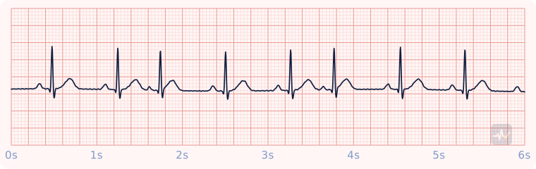
How to identify Premature atrial complexes (PACs)
- Rate
- That of the underlying sinus rhythm
- Rhythm
- Regular, interrupted by early beats
- P waves
- The early beat has a P that looks different, and it can hide in the T wave before it
- PR interval
- Normal, can be a little shorter on the early beat
- QRS
- Under 0.12 s
- Clincher
- An early, abnormally shaped P wave.
Rhythms it gets mistaken for
Premature atrial complexes (PACs) vs Unifocal PVCs
Premature atrial complexes (PACs) has an early P wave that looks different from the others. With unifocal PVCs you would see a P wave before each normal beat and no P wave in front of the early beats. It also has a narrow QRS, under 0.12 s, while unifocal PVCs would show narrow beats with wide early beats mixed in. A narrow QRS means the signal came from above the ventricles. The early beats start in the atria, not the SA node. How to spot unifocal PVCs: Early, wide beats that all look alike, with a T wave pointing opposite the QRS and a compensatory pause.
Premature atrial complexes (PACs) vs Sinus arrhythmia
Premature atrial complexes (PACs) has an early P wave that looks different from the others. With sinus arrhythmia you would see one upright P wave before every QRS, all the same shape. It also has a regular rhythm interrupted by early beats, while sinus arrhythmia would show a rate that speeds up and slows down in a smooth, repeating wave. The early beats start in the atria, not the SA node. How to spot sinus arrhythmia: The rate speeds up and slows down with breathing.
Premature atrial complexes (PACs) vs Atrial fibrillation
Premature atrial complexes (PACs) has an early P wave that looks different from the others. With A-fib you would see no P waves, only a wavy, chaotic baseline. It also has a regular rhythm interrupted by early beats, while A-fib would show an irregular rhythm with no pattern to the R-R intervals. How to spot A-fib: Irregularly irregular: no two R-R intervals match.
Treatment by certification level
Open your level. Treat the patient, not the strip.
Paramedic
No rhythm treatment needed.
- Treat the patient in front of you, not the strip.
AEMT
Pulse present and stable.
- Oxygen only if hypoxic or short of breath, IV access, monitoring.
- Acquire and transmit a 12-lead ECG. Interpreting it is a Paramedic skill.
- Transport and reassess. Request a paramedic if the patient worsens.
EMT
Pulse present: assess and support.
- Treat the patient, not the rhythm name: airway, breathing, circulation, vital signs.
- Oxygen if the SpO2 is under 94%, the patient is short of breath, or shows signs of shock.
- Request ALS and transport promptly. Reassess on the way.
An AED goes only on a patient who is unresponsive, not breathing and pulseless. It cannot feel a pulse, so on a patient with a pulse it can advise a shock that would do harm.